Describe the Layers of the Heart Wall
The wall of the heart is composed of three distinct layers. Human heart is situated in the space of thoracic cavity called mediastinum present between two lungs.

The Three Layers Of The Heart Wall Location Structure Function Video Lesson Transcript Study Com
The heart chamber is the space inside the heart and thats normally filled with blood.

. Cardiovascular System Learning Objectives Describe the three layers of the heart. View Test Prep - BIO 212 Exam 3 Study Guide part 2 from BIO 212 at North Carolina State University. Myocardium- Middle muscular layer forming atria and ventricles.
Describe the three major layers of the heart wall and how they relate to the pericardium. Want to see this answer and more. A Fibrous pericardium.
The epicardium which corresponds to the visceral pericardium protects the heart by reducing friction. The myocardium is composed of. It lines the cavities and valves of the heart.
The wall of the heart consists of three layers. It preforms the function of pumping what is necessary for the circulation of blood. All this red stuff is heart muscle called myocardium and then theres a membrane surrounding it and we call it the pericardium.
Describe the pathway of blood through the heart and lungs starting at the right atrium and ending at the aorta. The Layers of the Heart Wall The heart wall is composed of connective tissue endothelium and cardiac muscle. Describe the structure and function of each of the three layers of the heart wall.
Endocardium The innermost layer of the cardiac wall is known as the endocardium. This is the middle layer of the heart that contains the cardiac muscular tissue. It lines the cavities and valves of.
Each made up of different cells and all pertaining to different purposes that aid in the function of the heart. The heart wall consists of three layers. The inner layer of the heart.
Solutions for Chapter 13 Problem 4P. Median response time is 34 minutes for paid subscribers and may be longer for promotional offers. Answer to Describe the layers of the heart wall.
The heart wall is divided into three layers. Identify the three layers of arteries and veins. Epicardium myocardium and endocardium.
Check_circle Expert Answer thumb_up thumb_down Introduction. Describe the layers of the heart wall. Visceral layer of serous pericardium.
There arefenestrations openings in the epithelial cells of capillary walls. Layers of the Pericardium Heart Wall and Spiral Arrangement. The outer layer of the heart wall is the epicardium the middle layer is the myocardium and the inner layer is the endocardium.
The layers of the heart are as follows. Outer layer of the heart and inner most layer of the serous pericardium. Name and describe each of the heart valves.
The muscular middle layer of the wall of the heart. Name the major coronary arteries and the myocardial regions that they each supply. Loose Leaf Version of Holes Human Anatomy Physiology with Connect Plus 2 Semester Access CardLearnSmartAPR PhILS Online Access 13th Edition Edit edition.
Answer 1 Coverings and layers of the heart wall Pericardium is the outer covering of the heart. The wall of the heart is a thick layer and can be three layers of the heart wall that comprises this wall. In this picture above weve sliced the heart.
The esophageal wallincludes a middle layer of dense irregular connective tissue. The epicardium external layer the myocardium middle layer and the endocardium inner layer. An outer epicardium a middle myocardium and an inner endocardium.
It is mesodermal in origin and is slightly tilted to the left side of the body. The layers of the heart wall can be divided into three layers as can be seen clearly from the image below. Structurally the endocardium is comprised of loose connective tissue and simple.
It is the cardiac muscle that enables the heart to contract and allows for the synchronization of the heartbeat. The outer layer of the wall of the heart. The muscles of thethigh are composed of skeletal muscle tissue.
The Heart Wall Endocardium. Epicardium- Covers surface of the heart. Holes Essentials of Human Anatomy and Physiology 13th Edition Edit edition This problem has been solved.
Endocardium- Covers inner surfaces of heart including valves Describe the structure and functions of the four heart chambers. Experts are waiting 247 to provide step-by-step solutions in as fast as 30 minutes. Composed of mesothelium and adipose tissue.
Pericardium or the layer surrounding the heart Epicardium the outside layer of the heart Myocardium the middle layer of the heart Endocardium the innermost layer of the heart. Response times may vary by subject and question complexity. Describe the layers of the heart wall.
4 rows Key facts about the layers of the heart. There are two type of pericardium. This is the outermost layer of the heart and is one of the two layers of the pericardium.
The innermost layer of the cardiac wall is known as the endocardium. The walls of bloodcapillaries are composed of a thin epithelium. The heart wall The heart wall itself can be divided into three distinct layers.
Epicardium external layer myocardium middle layer and endocardium inner layer. The subendocardial layer lies between and joins the endocardium and the myocardium. The outermost covering of heart is fibrous pericardium that protects the heart and it also h View the full answer.

Heart Anatomy Anatomy And Physiology I
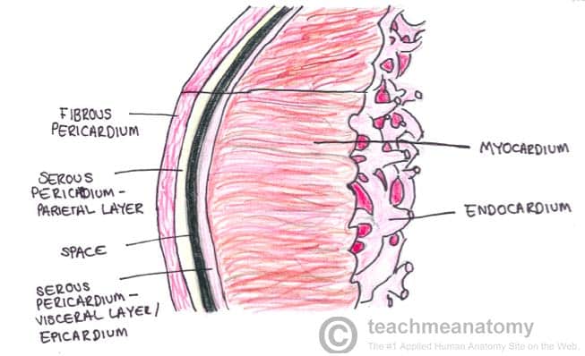
The Heart Wall Teachmeanatomy
Layers Of The Heart Flashcards Quizlet
No comments for "Describe the Layers of the Heart Wall"
Post a Comment